Healthcare providers are increasingly adopting the AI Fundus Camera as part of modern retinal screening programs. While traditional fundus cameras have been a trusted diagnostic tool for decades, advances in artificial intelligence have transformed how retinal images are captured, analyzed, and used to support clinical decision-making. An AI Fundus Camera combines high-quality retinal imaging with automated image analysis, helping providers identify eye diseases faster, improve workflow efficiency, and expand access to preventive eye care.
What Is an AI Fundus Camera?
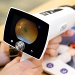
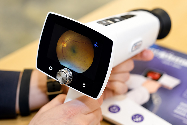
An AI Fundus Camera is a retinal imaging device that captures detailed photographs of the retina while using artificial intelligence algorithms to analyze images for signs of disease. Unlike traditional systems that rely solely on manual review by eye care professionals, AI-powered platforms provide automated screening support and near real-time diagnostic insights.
BeamMed’s AI Fundus Camera is designed to help healthcare organizations streamline retinal screening while supporting early disease detection and preventive care initiatives.
How Traditional Fundus Cameras Work
Traditional fundus cameras capture images of the retina, optic nerve, macula, and retinal blood vessels. These images are then reviewed by ophthalmologists, optometrists, or retinal specialists who evaluate them for abnormalities.
While traditional fundus imaging remains highly valuable, the process depends on:
- Manual interpretation by trained clinicians
- Availability of specialists to review images
- Potential delays between image capture and diagnosis
- Variability in interpretation based on experience and workload
In busy healthcare environments, these factors can impact screening efficiency and patient throughput.
AI Fundus Camera vs Traditional Fundus Camera: Key Differences
1. Automated Image Analysis
The biggest distinction between an AI Fundus Camera and a traditional fundus camera is the ability to automatically analyze retinal images.
Traditional systems require human review for every image. AI-powered systems evaluate retinal photographs against extensive datasets trained to recognize retinal abnormalities and disease patterns.
This technology can help identify potential concerns that require further clinical evaluation.
2. Faster Results
Traditional workflows often involve image capture followed by later review from a specialist. Depending on staffing and patient volume, results may take hours or days.
An AI Fundus Camera can provide screening results within seconds or minutes, allowing clinicians to discuss findings during the same patient visit. This significantly improves workflow efficiency and patient engagement. :contentReference[oaicite:0]{index=0}
3. Enhanced Screening Consistency
Human interpretation naturally varies among clinicians based on experience, training, and workload. AI algorithms provide standardized analysis using the same evaluation criteria for every image.
This consistency helps reduce variability and supports more uniform screening outcomes across multiple providers and locations.
4. Increased Accessibility
Traditional retinal screening programs often require access to ophthalmologists or retinal specialists.
AI-powered systems make retinal screening more accessible by enabling screening programs in:
- Primary care clinics
- Endocrinology practices
- Community health centers
- Rural healthcare facilities
- Population health programs
This expanded accessibility can help identify at-risk patients earlier in the disease process.
Benefits of an AI Fundus Camera
Improved Early Disease Detection
Many retinal diseases progress silently before symptoms become noticeable. Early identification is critical because treatment outcomes are generally better when intervention occurs sooner.
An AI Fundus Camera can evaluate thousands of image data points and identify subtle retinal changes that may indicate conditions such as:
Diabetic Retinopathy
Diabetic retinopathy remains one of the leading causes of vision loss among adults. AI-assisted retinal screening helps healthcare providers identify retinal damage earlier and improve screening compliance. :contentReference[oaicite:1]{index=1}
Age-Related Macular Degeneration
AI analysis can assist in detecting retinal changes associated with age-related macular degeneration, supporting timely referral and management.
Glaucoma Risk Indicators
Retinal imaging plays an important role in identifying optic nerve changes associated with glaucoma risk. AI-powered systems can assist providers by highlighting suspicious findings for further evaluation. :contentReference[oaicite:2]{index=2}
Workflow Efficiency for Healthcare Organizations
Healthcare systems are under constant pressure to improve efficiency while maintaining high-quality patient care.
An AI Fundus Camera helps streamline clinical operations by:
Reducing Specialist Burden
Automated analysis allows specialists to focus attention on patients who require further evaluation rather than manually reviewing every screening image.
Supporting Same-Day Decision Making
Immediate screening results enable clinicians to discuss findings with patients during the appointment, improving care coordination and reducing delays.
Enhancing Population Health Programs
Organizations focused on preventive care and chronic disease management can use AI-powered retinal screening to increase screening rates and improve patient outreach efforts.
For providers focused on quality measures and preventive care, technologies featured within BeamMed’s HEDIS solutions can support broader population health initiatives.
Why Healthcare Providers Are Moving Toward AI-Powered Retinal Screening
The healthcare industry continues to embrace technologies that improve accuracy, efficiency, and patient outcomes.
Several factors are driving adoption of the AI Fundus Camera:
Growing Demand for Preventive Care
Preventive screening programs are becoming increasingly important as healthcare organizations focus on earlier intervention and chronic disease management.
Healthcare Workforce Challenges
Many regions face shortages of eye care specialists. AI-assisted screening helps extend screening capabilities without requiring additional specialist resources. :contentReference[oaicite:3]{index=3}
Digital Healthcare Transformation
AI-enabled diagnostic tools align with broader healthcare trends toward automation, clinical decision support, and data-driven care delivery.
Providers interested in advanced diagnostic technologies can explore additional innovations through BeamMed’s technology solutions and clinical resources available within the BeamMed library.
The Future of AI Fundus Camera Technology
Artificial intelligence continues to evolve rapidly within ophthalmology and preventive healthcare. Future AI Fundus Camera systems are expected to provide even greater diagnostic capabilities, expanded disease detection, improved integration with electronic health records, and enhanced support for telemedicine programs.
As screening programs continue to grow, AI-powered retinal imaging will play a vital role in helping healthcare providers identify vision-threatening conditions earlier while improving operational efficiency.
Partner with BeamMed for Advanced AI Fundus Camera Solutions
BeamMed Inc. is a leader in innovative diagnostic technologies designed to improve preventive care and clinical outcomes. Through advanced retinal imaging solutions, including its AI Fundus Camera platform, BeamMed helps healthcare organizations enhance retinal screening programs, improve patient access to care, and support early disease detection initiatives.
With expertise in diagnostic imaging, population health solutions, HEDIS-focused screening programs, and healthcare technology integration, BeamMed provides the tools and support organizations need to implement efficient, scalable retinal screening programs. Whether serving primary care practices, endocrinology clinics, community health centers, or large healthcare systems, BeamMed delivers expert guidance and advanced technology solutions that help providers achieve better outcomes for their patients.
Frequently Asked Questions
Can an AI Fundus Camera identify diabetic retinopathy?
Yes. Modern AI Fundus Camera systems are specifically designed to detect signs of diabetic retinopathy and help providers identify patients who require additional ophthalmic evaluation.
Does an AI Fundus Camera replace an ophthalmologist?
No. AI serves as a clinical decision-support tool. Final diagnosis and treatment decisions remain the responsibility of qualified healthcare professionals.
How quickly does an AI Fundus Camera provide results?
Many AI-powered retinal screening systems can generate screening results within seconds, allowing clinicians to discuss findings during the same patient visit.
Can primary care clinics use an AI Fundus Camera?
Yes. AI-powered retinal imaging systems are increasingly used in primary care settings, endocrinology clinics, and community health programs to expand access to retinal screening services.