Advances in medical imaging technology are transforming how eye care providers identify and monitor retinal conditions. Among the most significant developments is the emergence of the AI Fundus Camera, a device that combines high-resolution retinal imaging with artificial intelligence analysis. Compared to traditional fundus imaging systems, AI-powered solutions offer faster assessments, improved consistency, and the ability to detect subtle abnormalities that may otherwise go unnoticed.
As healthcare systems prioritize early intervention and preventive care, understanding the differences between conventional imaging and AI-enhanced screening is essential for clinics, hospitals, and vision care centers seeking to improve outcomes while optimizing workflow efficiency.
How traditional fundus imaging works
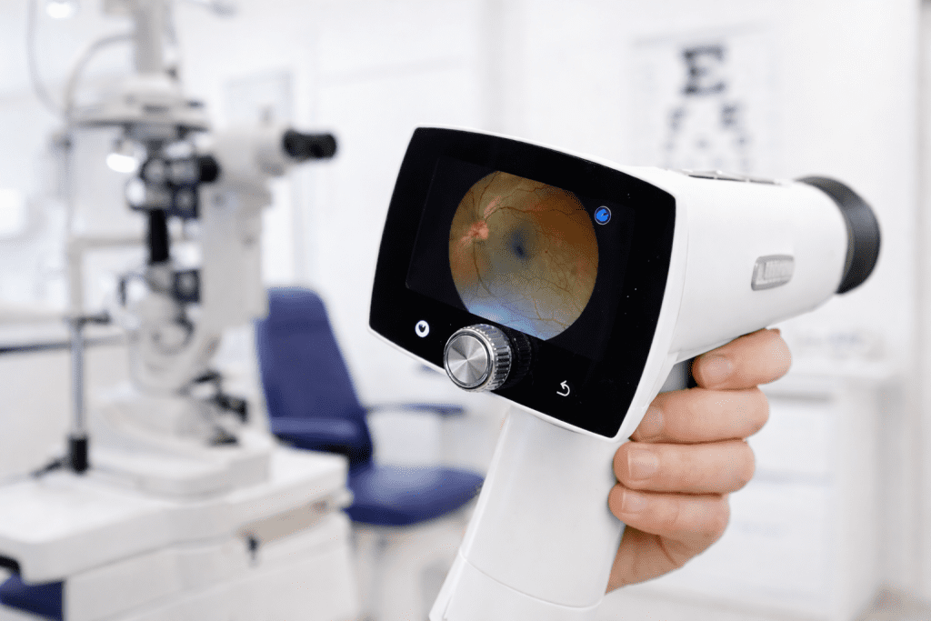
Traditional fundus cameras capture detailed photographs of the retina, optic nerve, and blood vessels at the back of the eye. These images are then interpreted by trained clinicians who look for signs of disease, structural changes, or abnormalities. While this method has been the standard for decades, it depends heavily on human interpretation, which can vary based on experience, fatigue, and workload.
Manual review processes can also slow down patient throughput, especially in high-volume settings. Clinics may face delays between imaging and diagnosis, particularly when specialists must review images remotely or during scheduled sessions.
What makes an AI Fundus Camera different
An AI Fundus Camera integrates advanced image analysis algorithms that evaluate retinal photographs in near real time. Instead of relying solely on manual interpretation, the system compares captured images against large datasets of annotated retinal patterns to identify potential concerns quickly and consistently.
Key capabilities that distinguish AI-powered systems include:
- Automated image analysis that highlights suspicious features
- Immediate feedback to clinicians during the patient visit
- Standardized evaluations that reduce variability between reviewers
These features allow providers to move from reactive care toward proactive screening, improving the likelihood of detecting issues before symptoms progress.
Improving early disease detection through AI
Early detection is critical for many retinal conditions, where treatment outcomes are significantly better when intervention occurs sooner rather than later. Traditional imaging relies on a clinician’s ability to identify subtle patterns visually, which may be challenging in early stages.
AI-driven systems enhance sensitivity by analyzing thousands of data points across each image, including micro-changes in vascular structure, tissue coloration, and anatomical patterns. This level of analysis can flag concerns that appear normal to the human eye, prompting further evaluation.
AI Fundus Camera technology also supports population-level screening programs, enabling primary care clinics and community health centers to identify at-risk patients who might otherwise lack access to specialized ophthalmic services.
Workflow efficiency and patient throughput
Beyond diagnostic advantages, AI-enabled imaging significantly improves operational efficiency. Traditional workflows often involve capturing images, storing them, scheduling review, and communicating results later. AI integration compresses this timeline by providing immediate insights.
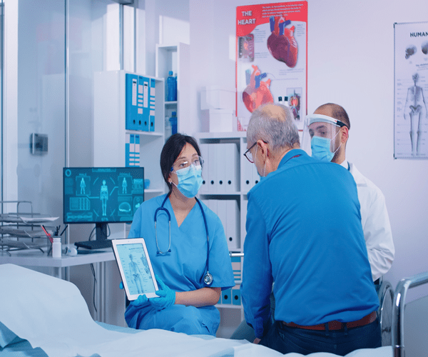
Clinics can discuss findings with patients during the same appointment, reducing follow-up visits and improving care continuity. This streamlined approach benefits both providers and patients by saving time and reducing uncertainty.
Consistency and reduction of human error
Human interpretation remains essential, but variability is inevitable. Factors such as experience level, workload, and environmental conditions can influence how images are assessed. AI systems provide a standardized baseline analysis, ensuring that no image is overlooked or misclassified due to fatigue or subjective judgment.
- Consistent evaluation criteria across all patients
- Reduced risk of missed early indicators
- Decision support for clinicians rather than replacement
This combination of automation and expert oversight strengthens clinical confidence and supports evidence-based decision-making.
Accessibility and expansion of screening services
Traditional retinal imaging systems often require specialized operators and dedicated ophthalmology settings. AI-powered devices are designed to be more user-friendly, enabling trained staff in primary care environments to capture high-quality images.
This expanded accessibility allows screening to occur in locations where specialist services may be limited, such as rural clinics, community health centers, or mobile medical units. By bringing advanced imaging closer to patients, healthcare providers can reach populations that might otherwise delay care.
Cost considerations and long-term value
While AI-enabled systems may involve higher initial investment, their long-term value can be substantial. Earlier detection can reduce the need for complex interventions later, lowering overall healthcare costs and improving quality of life for patients.
Operational savings also come from increased efficiency. Clinics can serve more patients per day, reduce administrative burden, and minimize repeat visits caused by delayed results.
Future-ready technology for modern healthcare
Healthcare is rapidly moving toward data-driven decision support and preventive care models. An AI Fundus Camera aligns with this shift by combining imaging, analytics, and digital integration into a single platform. As AI algorithms continue to evolve, these systems will become even more accurate and capable of identifying a broader range of conditions.
Organizations adopting AI-powered screening today position themselves at the forefront of innovation, offering advanced care while maintaining operational flexibility.
Why healthcare providers choose BeamMed
BeamMed delivers advanced medical imaging solutions designed to improve clinical performance, efficiency, and patient outcomes. By offering state-of-the-art AI-enabled technologies such as the AI Fundus Camera, BeamMed helps clinics integrate modern screening capabilities into everyday practice. Their solutions are supported by training, technical assistance, and implementation guidance, ensuring providers can maximize the value of their investment while delivering high-quality care. Call 800-769-6808
Frequently Asked Questions
How does an AI Fundus Camera improve screening accuracy?
AI algorithms analyze retinal images using pattern recognition trained on large datasets, helping identify subtle abnormalities that may be difficult to detect manually.
Can AI replace ophthalmologists?
No. AI systems act as decision-support tools, assisting clinicians by highlighting potential concerns while final interpretation remains with qualified professionals.
Is AI-powered retinal imaging suitable for primary care clinics?
Yes. Many AI fundus systems are designed for use outside specialized eye centers, enabling broader screening access.
How quickly are results available?
Most AI-enabled systems provide feedback within seconds to minutes, allowing clinicians to discuss findings during the same visit.


