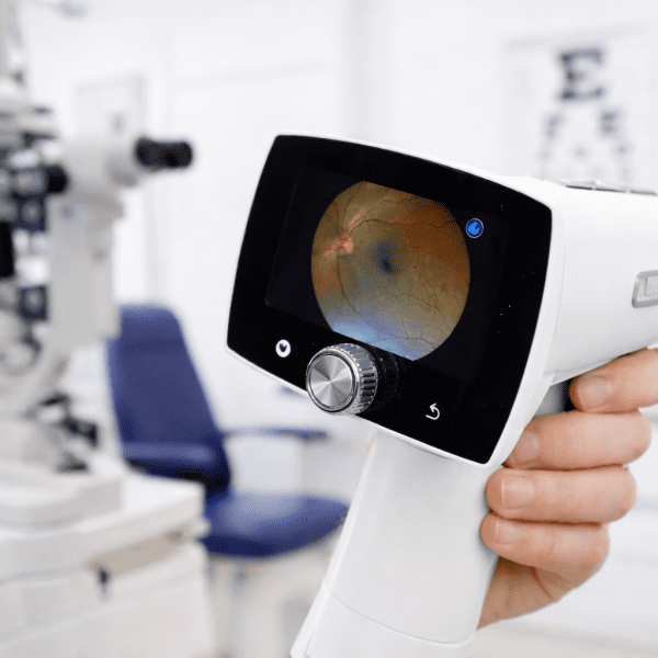

Preventive care works best when clinicians can identify health risks before symptoms interfere with a patient’s quality of life. Preventive screening technology can support that goal by bringing efficient, noninvasive assessments into primary care. Two complementary tools—retinal imaging and bone density assessment—give clinicians additional insight into eye health, systemic risk indicators, and skeletal health during routine care.
Used together, these technologies can help primary care teams build a more complete picture of a patient’s health while identifying when further evaluation or specialist referral may be appropriate.
How Preventive Screening Technology Supports Primary Care
Primary care providers are often the first to recognize early signs of chronic disease. However, limited appointment time, access barriers, and the absence of obvious symptoms can make some conditions difficult to identify at an early stage.
Preventive screening technology expands the information available during routine visits. Retinal imaging creates detailed images of the back of the eye, while bone density assessment estimates skeletal strength and potential fracture risk. Neither screening replaces a complete clinical evaluation, but each can provide useful information for determining appropriate next steps.
Understanding Retinal Imaging
Retinal imaging uses a specialized fundus camera to photograph structures at the back of the eye, including the retina, optic disc, macula, and visible blood vessels. These images may help trained professionals recognize findings that warrant closer evaluation.
An AI-supported fundus camera can assist with image acquisition and analysis, depending on the device and its intended use. The resulting information can help clinicians determine whether a patient should receive a comprehensive eye examination or be referred to an ophthalmologist or optometrist.
Retinal screening may be particularly relevant for people with diabetes, hypertension, age-related risk factors, or limited access to regular eye care. Abnormal findings should always be interpreted within the patient’s broader clinical history rather than treated as a stand-alone diagnosis.
Understanding Bone Density Assessment
Bone density assessment helps evaluate skeletal health and identify patients who may be at increased risk of osteoporosis or fractures. Although bone loss is often associated with aging, it can also be influenced by family history, low body weight, smoking, medication use, nutrition, hormonal changes, and certain medical conditions.
Portable technologies such as the MiniOmni bone density scanner can make skeletal assessment more accessible in primary care and community settings. Depending on the technology, measurements may be taken at peripheral skeletal sites rather than the hip or spine.
Peripheral assessment can support risk evaluation, but it is not necessarily interchangeable with central dual-energy X-ray absorptiometry, commonly called DXA. Results that indicate elevated risk may lead to additional testing, lifestyle guidance, medication review, or a referral for further evaluation.
Benefits of Combining the Two Screenings
Retinal and bone assessments examine different parts of the body, yet they share an important purpose: identifying risk before a serious complication occurs. Incorporating both into a preventive workflow may offer several practical benefits:
- Broader risk visibility: Clinicians can gather information related to eye and skeletal health during the same care pathway.
- Earlier follow-up: Concerning findings can prompt confirmatory testing or specialist referrals.
- Greater accessibility: Point-of-care technology may reduce the need for a separate initial screening appointment.
- Better patient engagement: Images and assessment results can help clinicians explain risks and recommended next steps.
These screenings are most valuable when they complement—not replace—medical history, physical examination, laboratory testing, established screening guidelines, and clinical judgment.
Building an Effective Primary Care Workflow
Successful implementation requires more than placing devices in an examination room. Practices should define which patients qualify for screening, who will operate the equipment, how results will be reviewed, and when referrals are necessary.
A structured workflow may include:
- Identifying eligible patients through age, medical history, medication use, or other risk factors.
- Explaining the purpose and limitations of each screening before testing.
- Capturing images or measurements according to device instructions.
- Documenting results in the patient’s health record.
- Creating clear pathways for repeat screening, confirmatory testing, and specialist care.
Primary care organizations should also consider staff training, patient privacy, device maintenance, quality assurance, and applicable regulatory requirements. Practices exploring implementation can review additional preventive screening resources when planning their approach.
Choosing Appropriate Screening Technology
Technology should fit the practice’s patient population and clinical objectives. Important considerations include ease of operation, portability, examination time, result presentation, staff training requirements, and the ability to integrate findings into existing workflows.
Clinicians should also understand what a device measures, its intended use, and the limitations of its output. A screening result should not create false reassurance or unnecessary concern. Clear patient communication is essential, especially when a result requires confirmation through a comprehensive eye examination, DXA scan, laboratory testing, or specialist consultation.
BeamMed Preventive Screening Solutions
BeamMed Inc. provides technologies for retinal imaging and bone density assessment, including an AI fundus camera and the MiniOmni bone density scanner. These products are intended to help healthcare organizations incorporate accessible screening capabilities into clinical workflows.
Primary care practices can visit the BeamMed website to learn more about available technology and determine whether these solutions align with their screening goals. When selected carefully and used within an appropriate clinical pathway, BeamMed’s products can support a reliable, patient-focused approach to preventive assessment.
Frequently Asked Questions
Can retinal imaging detect health problems outside the eye?
Retinal images may reveal vascular or structural findings associated with certain systemic conditions. However, these findings are not a stand-alone diagnosis and must be reviewed alongside the patient’s medical history and other clinical information.
Is a portable bone density screening the same as a DXA scan?
Not necessarily. Portable devices may assess peripheral skeletal sites using a different measurement method. A concerning result may lead a clinician to recommend central DXA or another confirmatory assessment.
How often should patients receive these preventive screenings?
Screening frequency depends on age, health history, current risk factors, previous results, and professional guidelines. A healthcare provider should establish an individualized schedule and recommend follow-up when appropriate.
Why combine retinal imaging with bone density assessment?
The screenings evaluate different health concerns but support the same preventive objective. Together, they can help primary care teams identify potential eye and skeletal risks and determine whether further evaluation is needed.